Experimental Poisoning with Baccharis coridifolia (Asteraceae) in Goats
Intoxicación Experimental con Baccharis coridifolia (Asteraceae) en Cabras
Laura S. Aguirre1,2,4* , Rodrigo García-Prieto4 , Agustín Avellaneda-Cáceres1,2,3 , Diego M. Medina4
  , Luis A. Colque-Caro1,2
  & Juan F. Micheloud1,2,4
1.
Consejo Nacional de investigaciones Científicas y Técnicas (CONICET), Rivadavia
941 (A4400), Salta, Argentina.
colquecaro.luis@inta.gob.ar
- https://orcid.org/0000-0002-3821-4683
2. Área
de Salud Animal “Dr. Bernardo J. Carrillo” - IIACS -CIAP -Instituto Nacional de
Tecnología Agropecuaria (INTA), RN 68 Km 172 (A4403), Cerrillos, Salta,
Argentina. micheloud.juan@inta.gob.ar - https://orcid.org/0000-0001-8709-895X
3. Cátedra
Elementos de Reproducción y Sanidad Animal, Facultad de Ciencias Naturales,
Universidad Nacional de Salta, Av. Bolivia 5150 (A4408FVY), Salta, Argentina. avellaneda.agustin@inta.gob.ar
- https://orcid.org/0000-0003-4744-1551
4. Facultad
de Ciencias Agrarias y Veterinarias, Universidad Católica de Salta, Campo
Castañares s.n. (A4400), Salta, Argentina. diegomartinnicolas@hotmail.com -
https://orcid.org/0000-0003-3080-7609, rodrigo.garcia@lagalesa.com.ar
https://orcid.org/0009-0000-5466-5057
Author
for correspondence: aguirre.sabrina@inta.gob.ar -
https://orcid.org/0000-0001-5644-5795
Abstract
This work aimed to experimentally reproduce
Baccharis coridifolia poisoning in goats. Eight adult Saanen goats were divided into two groups, four received
an oral dose of 5 g/kg live weight of dried and ground
plant and the rest of them were used as a control group.
The animals were inspected every 2 hours and the observed clinical signs were restlessness, anorexia, tachycardia, tachypnea,
muscle spasms, colic, mild tympanism, ruminal
atony, and death. The main macroscopic alterations were observed in the digestive system, consisting of edema and diffuse congestion of the mucosa
of all pre- stomachs, abomasum, and intestines. Histopathological examination revealed especially degeneration, coagulative necrosis, ulceration, and detachment of the epithelium from both the rumen and reticulum.
B. coridifolia is one of the most important toxic plants in Argentina. Although many species
are known to be susceptible, studies on this subject are scarce
in goats, which gives this work a greater relevance.
Keywords: Digestive disorder; Prestomachs, Small ruminants; Toxic plants
Resumen
El objetivo de este trabajo
fue reproducir experimentalmente la intoxicación con Baccharis coridifolia en la especie caprina.
Para llevar a cabo el ensayo
se emplearon ocho caprinos adultos
de la raza Saanen,
cuatro de ellos recibieron una dosis oral de 5 g/kg de peso vivo de planta seca y molida.
Los animales fueron inspeccionados cada 2 horas,
los signos clínicos que manifestaron fueron
intranquilidad, anorexia, taquicardia, taquipnea, espasmos musculares, cólico, timpanismo leve, atonía ruminal y muerte.
Las principales alteraciones macroscópicas se observaron a nivel del aparato digestivo, consistiendo en edema y congestión difusa de la mucosa de los preestómagos, abomaso e intestinos. El examen histopatológico reveló principalmente degeneración, necrosis coagulativa, ulceración y desprendimiento de epitelio de revestimiento de rumen y retículo. B. coridifolia es una
de las plantas toxicas más
importantes de Argentina. Si bien son susceptibles muchas especies, en los caprinos
son escasos los estudios
al respecto lo que le da a este trabajo
mayor relevancia.
Palabras Clave:
Desordenes digestivos; Preestómagos; Rumiantes
menores; Plantas
tóxicas

Aguirre, L. S. García-Prieto, R., Avellaneda-Cáceres, A., Medina, D. M., Colque-Caro, L. A., & Micheloud, J. F. (2023). Experimental Poisoning with Baccharis coridifolia (Asteraceae) in Goats. Revista Ciencias Naturales, 1(2), 78–83.
Recibido: 23/4/2023 Aceptado: 17/7/2023 Publicado: 15/8/2023 Editora
Asociada: Paula Narváez
INTRODUCTION
Baccharis coridifolia is a toxic plant commonly known in
Argentina as “mío-mío”
or “romerillo”, and it is widely
distributed in the central and southern regions of the country,
Uruguay, Bolivia,
Paraguay, and south of Brazil (Budel & Duarte,
2007). The poisoning is more frequent
in cattle but has
also been described in sheep, goats,
horses, and pigs (Tokarnia
et al., 2012).
Macrocyclic trichothecenes were reported
to be the main toxic principles, and they seem to be produced
by soil fungi of the genus Myrothecium (Berreta, 1996; Riet-Correa
& Méndez, 2007).
The mycotoxins isolated
from B. coridifolia and therefore produced by Myrothecium were
roridines A, D, and E, verrucarines A and J, and mycotoxin A (Perusia & Rodríguez,
2004). The roots of the plant absorb the mycotoxins and incorporate them into their
system without altering
their structure (Berreta, 1996; Riet- Correa & Méndez, 2007). The effects caused
by these mycotoxins are the alteration of cell
membrane polarity and enzyme systems, immunotoxicity, and inhibition of protein synthesis. Additionally,
the toxic dynamics of macrocyclic trichothecenes have been used to
evaluate their potential anticancer, fungistatic, and virucidal activity. According to the strong tendency
of trichothecenes to inhibit protein synthesis, they are considered
extremely irritating to the skin and mucous membrane
(Lozano & Díaz, 2006).
All parts of the plant are considered toxic.
In decreasing order of toxicity are flowers, seeds,
leaves, stems, and roots (Riet-Correa
& Méndez, 2007). There are also differences
in terms of toxicity when referring
to the sex of the plants
since female plants are more toxic than male plants due to their higher
accumulation of macrocyclic trichothecenes (Lozano & Díaz, 2006).
Natural poisoning occurs when animals without any previous exposure
to the plant are introduced into an area where B. coridifolia is present (Barros,
1998; Riet-Correa & Méndez,
2007; Tokarnia et al.,
2012). Even though experimental poisoning
has been carried
out in cattle,
sheep (Tokarnia et al., 2012),
and rabbits (Döbereiner et al., 1976),
toxicity in goats has been poorly studied, and the scarce information dates to articles from the last century (Flores
& Houssay, 1917).
The purpose
of this work was to reproduce experimentally the poisoning by B. coridifolia in goats to prove the susceptibility
of this species and describe the clinical and pathological findings.
MATERIALS AND METHODS
The UCASAL (Universidad Católica de
Salta) and CICUAL (Animal Care and Use Committee) Number 8-18 approved the study protocol. The requirements of the Argentine Animal Protection Policy (Ley 14346) were always
fulfilled. Eight adult
Saanen crossbred goats weighing
50, 7 +/- 8 kg were divided into two groups of four animals each. The first group was poisoned, and the second group
was the control group.
Before starting,
a general and objective gastrointestinal examination of all the animals was carried
out to verify their health
status.
B. coridifolia samples were collected both in the vegetative stage and early blooming
and dried at room temperature for one week (Fig. 1). Subsequently, leaves
and stems were milled to a size of approximately 2.5 mm. Medicago sativa L. leaves and stems were subjected to the same drying
and milling process.
The first group was the treatment group (animals 1, 2, 3, 4), which was administered B. coridifolia in a single dose of 5 g of dry matter/kg to each animal
by an oro-esophageal tube. The control
group (animals 5, 6, 7, 8) was administered only M. sativa in a single dose of 5 g of dry matter/kg to each animal
also by an oro-esophageal tube. Afterward, both groups
were confined separately and were offered
alfalfa hay and water ad libitum. The general clinical
inspection was done every two hours. Animals from the treatment group died within 12 to 24 hours after the poisoning
and the control group animals were euthanized, all the animals were necropsied. Tissue samples were collected, preserved in 10% buffered formalin, and processed routinely for sectioning and microscopic study. Sections
were stained with hematoxylin and eosin.
Observations and images were taken using an Olympus
U-TV0.5XC-3; DP22 camera mounted on an Olympus
Cx41 conventional optical microscope.
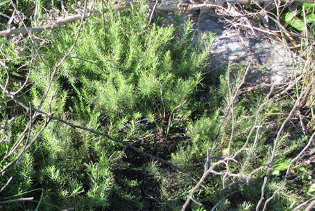
Figure 1. Baccharis coridifolia in the vegetative stage.
This variation
occurred from animal to
animal. Primarily shortening of the rumen papillae was observed
in all animals.
Rumen epithelial cells showed intense eosinophilic cytoplasm and karyorrhexis (animals 3 and 4). In addition, multiple
cells showed hydropic degeneration and several
Civatte’s corpuscles
(apoptosis). In some areas of the mucosa,
mild multifocal intercellular edema was identified causing a separation of the epithelial cells (Fig. 2B). Areas of necrosis
were identified in the papillae
covered by cellular debris and dense agglomerates of coccoid bacteria. In
the lamina propria, in one animal (4), an inflammatory infiltrate was observed
with the presence of intact and degenerated neutrophils.
Different degrees
of lymphocyte necrosis could be observed
in the thymus, spleen, and gut-associated lymphoid
tissue. Lymph node necrosis was also found, mainly involving the mesenteric lymph nodes, with moderate
necrosis of the follicles’ central
cells, showing
pyknosis and karyorrhexis or even with the loss of cellular detail and a large number of cellular debris. Hemorrhages were also observed (animal 2). In the liver, hepatocytes showed periportal tumefaction, vacuolization, and coagulative necrosis. was
observed in the four animals that died due to the intoxication.
DISCUSSION
This experiment confirmed that ingestion of B. coridifolia is
toxic for goats
as well as for other species, following
what has been described
in several works (Perusia et al., 2004; Rissi et al., 2005; Riet-Correa
& Méndez, 2007; Tokarnia
et al., 2012).
However, no information was found referring to experimental poisoning with B. coridifolia in goats or about toxic doses.
B. coridifolia is more
toxic during blooming in autumn, but poisoning
is more frequent in the spring (September–November) during its growth
period or vegetative stage when the plant shoots are mixed with grass
and appear to be more palatable and therefore
inadvertently ingested by animals (Barros,
1998; Rissi et al., 2005). In Northwestern Argentina, blooming
occurs during summer (January–March) when rains are more frequent.
The lethal dose of B. coridifolia for cattle varies between 0.25 to 0.5g/kg of dry matter in
the blooming period and 2g/kg of dry matter
during the vegetative
stage. (Tokarnia & Döbereiner, 1975; Varaschin et
al., 1998). Sheep are more resistant to poisoning and may have to ingest twice the toxic dose for cattle (Tokarnia & Döbereiner, 1975, Tokarnia
& Döbereiner, 1976). Conversely, no records referring to toxic doses in goats were found. In this work, the plant was toxic at a dose of 5g/ kg of dry matter to prove the susceptibility of this species. During blooming,
the toxic dose would probably be lower, because the toxicity of the plant increases during
this stage.
Animals in this experiment showed acute clinical signs strictly related
to gastrointestinal disorders as described in other papers,
both in goats (Barbosa
et al., 1994) and in other species
(Varaschin et al., 1998;
Rissi et al., 2005; Riet-Correa & Méndez, 2007; Pedroso et al., 2007; Tokarnia
et al., 2012).
The main lesions
produced by macrocyclic trichothecenes (roridine) identifiable
at necropsy include inflammatory lesions of the rumen, hepatitis, congestion, and pulmonary
edema (Radostits et al., 2002).
Changes in the prestomach, abomasum, and intestine
are the main macroscopic findings
in goats affected
by the disease. Variable
degrees of congestion, hemorrhage, edema, and mucosal
erosions are usually found
(Panziera et al., 2015). It is important
to emphasize that lesions in the
abomasum and segments of the small intestine
appear to be more severe in goats than in other species
(Barbosa et al., 1994). The histological findings
described in this work coincide
with those reported in other works
(Barbosa et al., 1994; Tokarnia
et al., 2012).
Lymphoid necrosis
seems to be a very consistent finding in
this poisoning (Varaschin et
al., 1998;
Rissi et al., 2005; Rozza et al., 2006) and some authors
report that this finding may be due to the toxicity
of this plant to B-lymphocytes (Rissi et al., 2005; Varaschin & Alessi, 2008).
Data
in the literature show a low prevalence or absence of cases of B. coridifolia poisoning in goats (Barbosa
et al., 1994; Pedroso et al., 2007; Tokarnia et al., 2012;
Rosa et al., 2013), contrary to what happens with cattle and sheep, which seem to be more susceptible to this poisoning
(Tokarnia et al., 2012). Due to the lack of cases described in goats, the register of cases of the Specialized Diagnostic Service
INTA-Salta has been taken
as a reference. According
to these data, poisoning outbreaks occurred in extensive systems
of mixed flocks (sheep and goats) grazing in areas contaminated by this plant when forage supply is scarce. In these cases, deaths occurred in both goats and sheep.
CONCLUSIONS
The susceptibility of goats to B. coridifolia poisoning was demonstrated even using the toxic
dose studied in sheep, and therefore a toxic dose of 5 g/kg of dry matter
could be confirmed for this species.
Thus, it was determined that the toxic dose for goats is higher than for cattle.
The clinicopathological findings of B. coridifolia poisoning in goats were also characterized and although there are some differences, the clinical picture
is similar to that of cattle and sheep reported by other authors.
ACKNOWLEDGMENTS
This study was supported by grants from Consejo de Investigación de la Universidad Católica de Salta (Proy. Res. 698/2020) and Instituto
Nacional de Tecnología Agropecuaria.
REFERENCES
Barbosa, J. D., Armién, A. G. & Tokarnia, C. H. (1994).
Intoxicação experimental
por Baccharis megapotamica var. weirii (Compositae) em ca- prinos. Pesquisa Veterinária Brasileira, 14, 5-13.
Barros, C. S. L. (1998). Livestock poisoning by Baccharis coridifolia.
En
T.
Garland & A. C. Barr (Eds), Toxic Plant and Other Natural
Toxicants (pp. 596-572).
CAB International.
Berreta, E. (1996). Malezas de campos sucios: El Mío-Mío. Boletín de Divulgación
del INIA, 60, 1-12. https://doi.org/10.4102/jsava.v70i1.753
Budel, J. M. & Duarte, M. R. (2007). Caracteres Morfoanatómicos de Partes Vegetativas
aéreas de Baccharis
coridifolia DC. (Asteraceae- Astereae). Latin American Journal of Pharmacy, 26 (5), 723-731.
http://sedici.unlp. edu.ar/handle/10915/7539
Döbereiner, J., Resende, A.
M. L.
& Tokarnia, C. H. (1976).
Intoxicação experimental por Baccharis coridifolia em coelhos. Pesquisa Agropecuária
Brasileira, 11 (9), 27-35.
Flores, C., Houssay, B. (1917). Estudios sobre el mio-mio,
nio o romerillo
(Baccharis coridifolia D.C.). Revista del Instituto Bacteriológico de Buenos Aires, 1, 59-100.
Lozano, M. C. & Díaz, G. J. (2006). Tricotecenos macrociclicos: toxinas hasta ahora no reconocidas en Colombia. Revista Colombiana de Ciencias Pecuaria. 19 (1), 46-60.
Panziera, W., Gonçalves, M. A., Lorenzett,
M.
P., Damboriarena, P., Argenta, F.
F., Laisse, J. M., Pavarini, S. P. & Driemeier, D. (2015). Intoxicação natural por Baccharis megapotamica var. weirii
em caprinos. Pesquisa Veterinária Brasileira35(4), 360-364. https:// doi.org/10.1590/S0100-736X2015000400008
Pedroso, P. M. O., Pescador, C. A., Oliveira, E. C., Sonne,
L., Bandarra, P. M., Raymundo, D. L. & Driemeier, D. (2007).
Intoxicações naturais por plantas em ruminantes
diagnosticadas no Setor de Patologia Veterinária da UFRGS no período de 1996-2005. Acta
Scientiae Veterinariae, 35(2), 213-218.
Perusia, O. R. & Rodríguez, R. (2004). Plantas toxicas y micotoxinas. Manual Divulgación Técnica Argentina, 4, 9-13.
Radostits, O. M., Gay, C. C., Blood, D. C. & Hinchcliff K. W. (2002).
Intoxicación por tricotecenos. En Medicina Veterinaria: tratado de
las enfermedades del ganado bovino,
ovino, porcino, caprino y equino (pp.2017-2019). Mc Graw-Hill-Interamericana.
Riet-Correa, F. & Méndez, M. C. (2007). Intoxicações por plantas e micotoxinas. En F. Riet-Correa, A. L Schild, Lemos, R. A. & Borges, J. R. (Eds.), Doenças de ruminantes e eqüídeos. Pallotti.
Rissi, D.
R., Rech, R. R., Fighera, R. A., Cagnini,
Q., Kommers, G. D. & Barros, C. S. L. (2005). Intoxicação espontânea por Baccharis coridifolia em bovinos. Pesquisa Veterinária Brasileira, 25, 111-114. https://doi.org/10.1590/ S0100-736X2005000200008
Rosa, F. B., Caprioli, R. A., Silva, T. M., Galiza, G.
J. N., Barros, C. S. L., Irigoyen, L. F., Fighera,
R.
A. & Kommers, G. D. (2013). Doenças de caprinos
diagnosticadas na região central do Rio Grande do Sul: 114 casos. Pesquisa Veterinária Brasileira, 33, 199-204.
https://doi.org/10.1590/ S0100-736X2013000200011
Rozza, D. B., Raymundo, D. L., Corrêa,A. M. R., Leal, J., Seitz, A. L., Driemeier, D. & Colodel, E. M. (2006). Intoxicação espontânea por Baccharis coridifolia (Compositae) em ovinos. Pesquisa Veterinária Brasileira, 26(1), 21-25. https://doi. org/10.1590/S0100-736X2006000100005
Tokarnia, C. H. & Döbereiner, J. (1975).
Intoxicação experimental em bovinos por “mio-mio”, Baccharis coridifolia. Pesquisa
Agropecuaria Brasileira, 10, 79-97.
Tokarnia, C. H. & Döbereiner, J. (1976). Intoxicação experimental em ovinos por “mio-mio”,
Baccharis coridifolia. Pesquisa Agropecuaria Brasileira, 11, 19-26.
Tokarnia, C. H., Farias Brito,
M., Barbosa J. D., Vargas Peixoto
P. & Döbereiner J. (2012). Plantas que afectam esencialmente o trato digestório: Bacccharis coridifolia. En Plantas tóxicas do Brasil para animais
de produção (pp. 95-102).
Helianthus.
Varaschin, M. S & Alessi, A. C. (2008). Histopathological examination of lymphoid organs in cattle and mice experimentally poisoned by Baccharis coridifolia: Immunohistochemical characterization of B and T lymphocytes. Brazilian Journal of Veterinary
Research and Animal Science, 45(4),
277-283. https://doi.org/10.11606/ issn.1678-4456.bjvras.2008.26686
Varaschin, M. S., Barros, C. S. L. & Jarvis, B. B. (1998). Intoxicação experimental por Baccharis coridifolia (Compositae) em bovinos. Pesquisa Veterinária Brasileira, 18(2), 69-74. https://doi. org/10.1590/S0100-736X1998000200006